

Laser Vision Correction technology is now more than half a century old.
1940’s – the Beginning
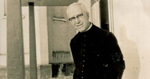
Figure 1 Father Waclaw Szuniewicz – pioneering refractive surgery
The idea of surgically altering the shape of the cornea to change the eyes’ focusing was first investigated in 1948 by Father Waclaw Szuniewicz in Poland and later at Yale University in the United States.
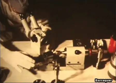
Figure 2. Dr Barraquers’ cryolathe setup for keratomileusis
1960’s
Professor Barraquer developed this further until 1964 when he described keratomileusis (Greek for sculpted cornea) and keratophakia (corneal lens).
Keratomileusis is really the no-laser form of laser vision correction. A layer of cornea is removed, reshaped and re-attached to the cornea thus resulting in a new corneal shape and changed focusing power of the eye. This worked but was an order of magnitude less precise than laser vision correction. It did show us however what was possible, and it just took for the application of laser technology to make this the procedure we have today.
1970’s
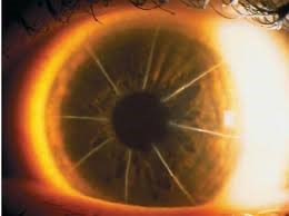
Figure 3. Radial Keratotomy cuts in the cornea
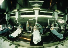
Figure 4. The production line system for Radial keratotomy
Fyodorov (a Russian ophthalmic surgeon) took a different approach and developed the technique of radial keratotomy.
The idea of cutting the cornea to cause selective flattening was not new having been described by a Japanese surgeon but Fyodorov quantified the effect of different size cuts in different locations on the cornea to give a predictable change in focusing. A diamond knife is used to make a series of radial cuts in the cornea to correct for myopia.
Interesting that one of the earliest applications of Excimer Laser to refractive surgery was to use the laser (Meditec) to make the cuts for radial keratotomy (rather than the diamond knife).
1980’s – Excimer Laser Technology Developed
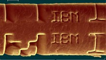
Figure 5. ‘IBM’ etched into a human hair with the excimer laser
Cutting the cornea with radial incisions is not the best way to correct refractive error and Dr Marshall (London) recognised in the 1980s that the cornea needed to be removed in the shape of a disc (as was performed in the earlier technique of keratomileusis) thus introducing PRK.
Excimer Laser technology was investigated and developed at IBM in the early 1970’s. The excimer laser uses reactive gases such as Chlorine and fluorine mixed with inert gases such as Argon, Krypton and Xenon which, when electrically excited, emit energetic pulses of Ultraviolet light. Pulses of this high-energy light can be used to precisely reshape materials such as polymers and corneal tissue.
1990’s – Precision of the Excimer Laser
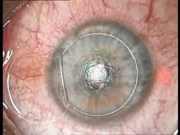
Figure 6. Lasik flap lifted from the cornea before the excimer laser is used to reshape the underlying cornea
Collaboration between Dr. Trokel, photo chemist – R. Srinivasan and materials scientist – J. Wynne resulted in the application of the precision of the Excimer Laser to corneal corrective surgery. PRK was first performed on a human eye in Dresden 1986 by Dr Seiler. Dr Trokel followed soon after in 1987 and holds the patents for Excimer Laser Vision Correction.
PRK remains an excellent treatment choice for the correction of refractive error today.
LASIK makes the corneal reshaping procedure better for patients. LASIK is a 2 step procedure where a corneal flap is raised to preserve the surface layers of the cornea and then the underlying cornea is reshaped with the Excimer laser. The flap is replaced onto the cornea leaving the surface of the eye intact. Drs. Pallikaris (Greece) and Burrato (Italy) both contributed to the development of LASIK in 1989.
2010’s – Small Incision Lenticule Extraction
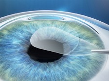
Figure 7. SMILE; a lenticule is created with a Femtosecond laser and removed through a keyhole incision
Most recently an entirely new approach to corneal refractive surgery has been introduced. The concept of refractive correction by removing a lenticule-shaped piece of corneal tissue is not new and really dates back to Dr Barraquer in the 1960s.
Using laser to create the lenticule however, makes this an accurate and desirable procedure. Zeiss began work on this concept in 2004 looking to apply Femtosecond laser technology (which was already in use for flap creation in LASIK) to an intrastromal lenticule for refractive correction.
The first SMILE (Small Incision Lenticule Extraction) procedure occurred in 2007 and since then the SMILE procedure has become a preferred option for the correction of myopia.
Laser Vision Correction Today
Laser vision correction surgery is able to treat short-sightedness (myopia), long-sightedness (Hyperopia) and astigmatism. With appropriate patient selection and attention to detail, it is a predictable and safe procedure that has helped more than 20 million people in the USA alone.